Actinic keratosis: how field-directed treatments compare
Actinic keratosis is a precursor to squamous cell carcinoma. What the progression data show, and how fluorouracil, imiquimod, and photodynamic therapy compared in a head-to-head trial.

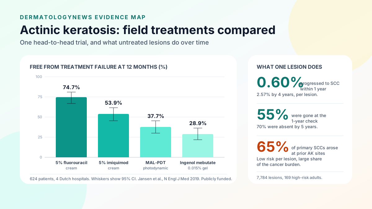
Actinic keratosis is the most common precancerous skin lesion, and it is a direct precursor of cutaneous squamous cell carcinoma (SCC). The risk that any single lesion progresses is small: in a cohort study that tracked 7,784 lesions on the face and ears of 169 high-risk participants, progression to primary SCC was 0.60 percent at 1 year and 2.57 percent at 4 years, and 55 percent of lesions were no longer present at 1 year [1]. The reason clinicians still treat is the reverse calculation from the same cohort: roughly 65 percent of all primary SCCs diagnosed arose at sites previously identified as actinic keratosis [1].
What actinic keratosis is, and what "field cancerization" means
An actinic keratosis is a rough, scaly patch that develops on skin with heavy cumulative ultraviolet exposure: the scalp in balding men, the forehead, the nose, the ears, the forearms, and the backs of the hands. It represents an early, confined disturbance of keratinocyte growth rather than an invasive cancer.
Field cancerization is the observation that these lesions rarely arrive alone. Where several visible actinic keratoses cluster in a continuous area, the surrounding skin that still looks normal often carries the same ultraviolet-driven changes below the threshold of visibility [4]. That observation is what motivates treating an entire area rather than only the spots a clinician can see and count.
The progression numbers, in both directions
The most-cited natural-history data come from the Veterans Affairs Topical Tretinoin Chemoprevention Trial, in which participants were examined every 6 months for up to 6 years with digital photographs used to track individual lesions across visits [1].
- Per-lesion progression is low. The risk that one clinically diagnosed actinic keratosis became a primary SCC, invasive or in situ, was 0.60 percent at 1 year and 2.57 percent at 4 years [1].
- Most lesions do not persist. 55 percent of the actinic keratoses followed clinically were absent at the 1-year visit, and 70 percent were absent at 5 years [1].
- Yet most SCCs came from them. About 65 percent of primary SCCs, and 36 percent of primary basal cell carcinomas, arose in lesions previously diagnosed clinically as actinic keratosis [1].
These findings sit together without contradiction. Any individual lesion is unlikely to become a cancer, and many resolve on their own, but across a sun-damaged field the aggregate contribution to keratinocyte cancer is substantial [1]. This cohort was drawn from a high-risk veteran population, so the absolute rates should not be read as applying to every patient.
How the field-directed options compared head to head
For years, guidelines gave no clear preference among field treatments [2]. A randomized trial published in the New England Journal of Medicine addressed that directly. It enrolled 624 patients at four Dutch hospitals, each with five or more actinic keratoses on the head within one continuous area of 25 to 100 square centimetres, and assigned them to 5 percent fluorouracil cream, 5 percent imiquimod cream, methyl aminolevulinate photodynamic therapy (MAL-PDT), or 0.015 percent ingenol mebutate gel [2].
The primary outcome was the proportion of patients with a reduction of 75 percent or more in lesion count, measured 12 months after treatment ended. At that point, the cumulative probability of remaining free from treatment failure was:
| Treatment | Free from treatment failure at 12 months | 95% CI |
|---|---|---|
| 5% fluorouracil cream | 74.7% | 66.8 to 81.0 |
| 5% imiquimod cream | 53.9% | 45.4 to 61.6 |
| MAL photodynamic therapy | 37.7% | 30.0 to 45.3 |
| 0.015% ingenol mebutate gel | 28.9% | 21.8 to 36.3 |
Compared with fluorouracil, the hazard ratio for treatment failure was 2.03 with imiquimod, 2.73 with MAL-PDT, and 3.33 with ingenol mebutate, all with P values of 0.001 or lower. No unexpected toxic effects were documented [2].
Two features make this trial unusually informative. It was funded by the Netherlands Organization for Health Research and Development rather than by a manufacturer, and it measured outcomes a full year after treatment rather than at the end of the treatment course, which is where short-term clearance figures tend to look most favourable [2].
Where photodynamic therapy fits
The Dutch trial placed one specific PDT protocol third of four, but that is a narrower finding than "PDT does not work." Other evidence describes what the treatment does under different conditions.
A phase III trial of BF-200 aminolevulinic acid with narrow-spectrum red light, conducted at seven German centres, followed patients who had achieved complete clearance. The probability that a participant was initially completely cleared and remained cleared for 12 months was 57.4 percent with BF-200 ALA versus 20.0 percent with placebo, and at the level of individual lesions, 85.4 percent versus 31.5 percent. Among participants with cosmetic impairment at baseline, 83.3 percent rated the 12-month cosmetic outcome as very good or good [4]. Four of the authors were employees of the manufacturer, Biofrontera Bioscience, which is disclosed in the paper.
A meta-analysis of 10 randomized trials, total sample 277, tested PDT combined with a topical agent against either treatment alone. Combination therapy improved participant complete clearance, relative risk 1.63 (95% CI 1.15 to 2.33), and lesion-specific clearance, relative risk 1.48 (95% CI 1.04 to 2.11). Partial clearance did not reach significance, relative risk 1.19 (95% CI 0.84 to 1.67). The authors judged the included studies to be at high risk of performance and detection bias, and noted that pain and local skin reactions were poorly reported [5].
Cosmetic outcome is part of why PDT is chosen. A prospective non-interventional study of 224 patients treated with methyl aminolevulinate artificial daylight PDT in Germany, median age 75, found that all five photoaging measures assessed, global photoaging score, mottled pigmentation, tactile roughness, telangiectasia, and fine lines, improved significantly from baseline at month 3, and 81.3 percent of patients rated the cosmetic result as good or very good [6]. That study had no control arm and was conducted by authors with Galderma affiliations, so it describes what patients and investigators observed rather than establishing a comparative benefit [6].
On seasonal timing
Scheduling field treatment for the lower-ultraviolet part of the year is a widespread clinical convention. The mechanism behind it is reasonable: field-directed treatment deliberately provokes an inflammatory reaction across the treated area, and skin in that state is temporarily more sensitive to sun.
The evidence base is quieter than the convention. None of the trials cited in this article compared treatment timing by season or tested whether starting in autumn produces better outcomes than starting in spring [1][2][4][5][6]. Daylight PDT protocols run in the opposite direction, since they depend on adequate outdoor light and comfortable temperature, which is one practical reason artificial daylight systems have been studied [6]. Readers who want the general case for year-round sun protection will find it in our photoprotection guide.
Adverse events, limitations, and realistic expectations
- Field treatment is meant to cause a reaction. Topical fluorouracil and imiquimod produce redness, crusting, erosion, and discomfort across the treated area for a period of weeks. That reaction is the mechanism, not a complication, but it affects work, appearance, and adherence, and it should be planned for rather than discovered.
- Adherence is part of the result. The Dutch trial's outcome measures reflect treatments used as directed under trial conditions, which is not automatically what happens at home over several weeks of visible inflammation [2].
- One protocol is not the whole modality. The trial tested MAL-PDT with a specific protocol. Different photosensitizers, light sources, and combination regimens produce different results [2][4][5].
- Sponsorship is uneven across the evidence. The head-to-head trial was publicly funded [2]. The BF-200 ALA follow-up included manufacturer employees among its authors [4], and the artificial daylight study was conducted with Galderma affiliations and no control arm [6]. These are not reasons to discard the findings, but they are reasons to weight the independent comparative data more heavily.
- Guideline quality varies. A systematic appraisal using the AGREE II and AGREE-REX instruments identified only three eligible actinic keratosis guidelines, from Canada, Germany, and the United Kingdom. Scores ranged from 65 to 92 percent for the German guideline down to 31 to 71 percent for the Canadian one, with stakeholder involvement and patient values identified as the weakest domains across the set [3].
- Treatment does not remove the underlying risk. Sun-damaged skin remains sun-damaged skin. Recurrence and new lesions are expected, and ongoing surveillance matters more than any single course.
- These are not cosmetic decisions. Actinic keratosis is a precursor lesion, and its management belongs in a clinical setting rather than in a treatment-menu comparison.
When it might be something else
A rough or scaly patch on sun-exposed skin has a differential that includes seborrheic keratosis, discoid lupus, and, importantly, lesions that have already progressed. A lesion that is tender, ulcerated, bleeding, rapidly enlarging, or thickened deserves prompt assessment rather than field treatment, because invasive SCC and basal cell carcinoma are managed differently [1]. Pigmented patches on sun-exposed skin raise a separate set of questions, covered in our guide to solar lentigines and our review of facial hyperpigmentation. Only clinical examination, and biopsy where indicated, resolves these distinctions.
Bottom line
Actinic keratosis carries a low per-lesion risk of becoming squamous cell carcinoma, 0.60 percent at 1 year in the most closely tracked cohort, while accounting for the majority of the SCCs that cohort produced [1]. When four field-directed treatments were compared directly over 12 months in 624 patients, 5 percent fluorouracil cream was the most effective of the four, keeping 74.7 percent of patients free from treatment failure, ahead of imiquimod, MAL-PDT, and ingenol mebutate [2]. Photodynamic therapy retains a role, with sustained 12-month clearance in a placebo-controlled trial and better clearance when combined with a topical agent, though that evidence is more industry-linked and more heterogeneous [4][5]. Seasonal scheduling is a sensible convention that trial evidence has not directly tested. What the data support most clearly is assessment by a clinician, treatment matched to how much of the field is involved, and continued follow-up afterwards.
Common questions
- Does an actinic keratosis always turn into skin cancer?
- No. In a cohort that tracked 7,784 individual lesions on the face and ears of 169 high-risk participants, the risk that a single actinic keratosis progressed to squamous cell carcinoma was 0.60 percent at 1 year and 2.57 percent at 4 years. Most lesions did not persist: 55 percent were gone at 1 year. The risk per lesion is low, but about 65 percent of the squamous cell carcinomas in that cohort arose at sites previously diagnosed as actinic keratosis, so the lesions still matter.
- What is the difference between lesion-directed and field-directed treatment?
- Lesion-directed treatment, such as cryotherapy, targets individual visible lesions. Field-directed treatment applies a cream, gel, or light-activated drug across a whole sun-damaged area, on the reasoning that the surrounding skin contains changes not yet visible. Which approach fits depends on how many lesions there are and how much of the area is affected, and that is a clinical judgement.
- Which field-directed treatment worked best in head-to-head testing?
- In a Dutch randomized trial of 624 patients with multiple lesions on the head, 5 percent fluorouracil cream was the most effective of the four treatments compared. At 12 months after treatment ended, 74.7 percent of the fluorouracil group remained free from treatment failure, versus 53.9 percent for imiquimod, 37.7 percent for methyl aminolevulinate photodynamic therapy, and 28.9 percent for ingenol mebutate.
- Is autumn the right time to treat sun damage?
- Scheduling field treatment for lower-ultraviolet months is a common clinical convention, and treated skin is temporarily inflamed and sun-sensitive. None of the trials cited here compared treatment timing by season, so the convention rests on mechanism and practicality rather than on trial evidence.
- How do I know whether a rough spot is an actinic keratosis or something else?
- You cannot reliably tell by looking. Actinic keratoses, seborrheic keratoses, early squamous cell carcinoma, and basal cell carcinoma overlap in appearance, and a lesion that bleeds, ulcerates, grows quickly, or feels tender needs assessment rather than a field cream. Diagnosis belongs with a dermatologist, and biopsy settles the uncertain cases.
References
- Criscione VD, Weinstock MA, Naylor MF, Luque C, Eide MJ, Bingham SF. Actinic keratoses: natural history and risk of malignant transformation in the Veterans Affairs Topical Tretinoin Chemoprevention Trial. — Cancer, 2009 · PMID: 19382202 · DOI: 10.1002/cncr.24284
- Jansen MHE, Kessels JPHM, Nelemans PJ, et al. Randomized trial of four treatment approaches for actinic keratosis. — New England Journal of Medicine, 2019 · PMID: 30855743 · DOI: 10.1056/NEJMoa1811850
- Wessely A, Steeb T, Heppt F, et al. A critical appraisal of evidence- and consensus-based guidelines for actinic keratosis. — Current Oncology, 2021 · PMID: 33617511 · DOI: 10.3390/curroncol28010093
- Reinhold U, Philipp-Dormston WG, Dirschka T, et al. Long-term follow-up of a randomized, double-blind, phase III, multi-centre study to evaluate the safety and efficacy of field-directed photodynamic therapy of mild to moderate actinic keratosis using BF-200 ALA versus placebo and the BF-RhodoLED lamp. — Journal of the European Academy of Dermatology and Venereology, 2025 · PMID: 39666443 · DOI: 10.1111/jdv.20452
- Heppt MV, Steeb T, Leiter U, Berking C. Efficacy of photodynamic therapy combined with topical interventions for the treatment of actinic keratosis: a meta-analysis. — Journal of the European Academy of Dermatology and Venereology, 2019 · PMID: 30710390 · DOI: 10.1111/jdv.15459
- Szeimies RM, Brückner M, Hoffmann M, et al. Photoaging and cosmetic result with artificial daylight photodynamic therapy using methyl aminolevulinate. — Acta Dermato-Venereologica, 2025 · PMID: 40263972 · DOI: 10.2340/actadv.v105.43245